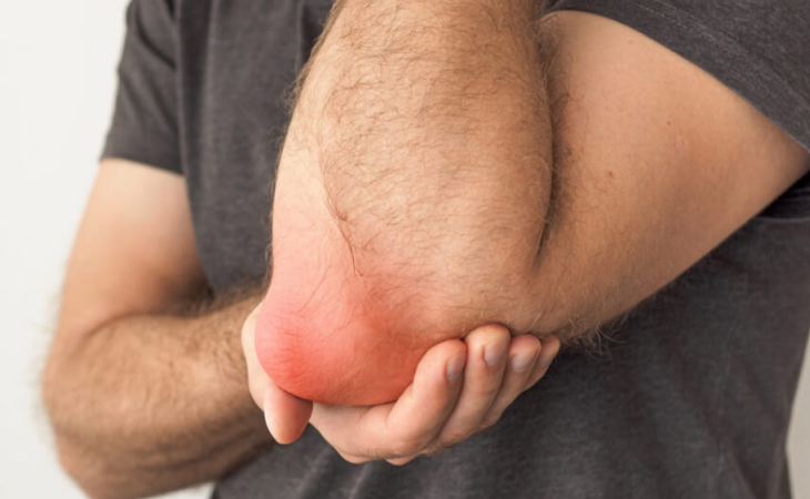
Bursitis
A bursa is a closed, fluid-filled sac that functions as a cushion and gliding surface to reduce friction between tissues of the body. The major bursae are located adjacent to the tendons near the large joints, such as in the shoulders, elbows, hips, and knees. When the bursa becomes inflamed, the condition is known as bursitis. Bursitis is usually a temporary condition. It may restrain motion, but generally does not cause deformity. The most common causes of bursitis are injury or overuse, although infection may also be a cause. Bursitis is also associated with other diseases, such as arthritis, thyroid disease, and diabetes.
symptoms of bursitis
The following are the most common symptoms of bursitis. However, each individual may experience symptoms differently
Bursitis can cause pain, localized tenderness, and limited motion. Swelling and redness may occur if the inflamed bursa is close to the surface (superficial). Chronic bursitis may involve repeated attacks of pain, swelling, and tenderness, which may lead to the deterioration of muscles and a limited range-of-motion. The symptoms of bursitis may resemble other medical conditions or problems. Always consult your physician for a diagnosis.
Risk factors for bursitis
Bursitis most often occurs in people who are in poor physical condition and/or have bad posture. Bursitis may also occur by overusing an affected limb, or by using an affected limb incorrectly
What are the different types of bursitis?
Although bursitis can occur anywhere in the body where bursae are located, there are several specific types of bursitis, including the following:
- Anterior Achilles tendon bursitis
This type of bursitis is also called Albert’s disease. Extra strain on the Achilles tendon, such as injury, disease, or shoes with rigid back support, causes this condition, which is characterized by inflammation of the bursa located in front of the attachment of the tendon to the heel.
- hip bursitis
Also called trochanteric bursitis, hip bursitis is often the result of injury, overuse, spinal abnormalities, arthritis, or surgery. This type of bursitis is more common in women and middle-aged and older people.
- knee bursitis
Bursitis in the knee is also called goosefoot bursitis or Pes Anserine bursitis. The Pes Anserine bursa is located between the shin bone and the three tendons of the hamstring muscles, on the inside of the knee. This type of bursitis may be caused by lack of stretching before exercise, tight hamstring muscles, being overweight, arthritis, or out-turning of the knee or lower leg.
- posterior Achilles tendon bursitis
This type of bursitis, also called Haglund’s deformity, is located between the skin of the heel and the Achilles tendon (which attaches the calf muscles to the heel). Aggravated by a type of walking that presses the soft heel tissue to the hard back support of a shoe, this type of bursitis occurs mostly in young women.s type of bursitis is also called Albert’s disease. Extra strain on the Achilles tendon, such as injury, disease, or shoes with rigid back support, causes this condition, which is characterized by inflammation of the bursa located in front of the attachment of the tendon to the heel.
- elbow bursitis
Elbow bursitis is caused by the inflammation of the olecranon bursa located between the skin and bones of the elbow. Elbow bursitis can be caused by injury or constant pressure on the elbow (for example, when leaning on a hard surface)
- kneecap bursitis
Also called prepatellar bursitis, this type of bursitis is common in people who sit on their knees a lot, such as carpet layers and plumbers.
How is bursitis diagnosed?
In addition to a complete medical history and physical examination, diagnostic procedures for bursitis may include the following:
x-ray – a diagnostic test which uses invisible electromagnetic energy beams to produce images of internal tissues, bones, and organs onto film.
computed tomography scan (Also called a CT or CAT scan) – a diagnostic imaging procedure that uses a combination of x-rays and computer technology to produce cross-sectional images (often called slices), both horizontally and vertically, of the body. A CT scan shows detailed images of any part of the body, including the bones, muscles, fat, and organs. CT scans are more detailed than general x-rays.
magnetic resonance imaging (MRI) – a diagnostic procedure that uses a combination of large magnets, radiofrequencies, and a computer to produce detailed images of organs and structures within the body.
arthrogram – an x-ray to view bone structures following an injection of a contrast fluid into a joint area. When the fluid leaks into an area that it does not belong, disease or injury may be considered, as a leak would provide evidence of a tear, opening, or blockage.
aspiration – involves a removal of fluid from the swollen bursa to exclude infection or gout as causes.
blood tests (to confirm or eliminate other conditions)
Treatment for bursitis:
- Specific treatment for bursitis will be determined by your physician based on:
- your age, overall health, and medical history
- extent of the condition
- your tolerance for specific medications, procedures, or therapies
- expectations for the course of the condition
- your opinion or preference
- The treatment of any bursitis depends on whether or not it involves infection.
aseptic bursitis - a non-infectious condition caused by inflammation resulting from local soft-tissue trauma or strain injury.
Treatment may include
- R.I.C.E. – Rest, Ice, Compression, and Elevation
- anti-inflammatory and pain medications such as ibuprofen or aspirin
- ultrasound – a diagnostic technique which uses high-frequency sound waves to create an image of the internal organs.
- aspiration of the bursa fluid for evaluation in the laboratory
- injection of cortisone into the affected area
- septic bursitis -bursa that becomes infected with bacteria
- antibiotic medications
- repeated aspiration of the inflamed fluid
- surgical drainage and removal of the infected bursa sac (bursectomy)
Quick Contacts
Please feel free to contact us for any medical inquiry
- Emergency call : +91 94274 18181
- Email : contact@trishahospital.com
- Location: Trisha multispeciality hospital, Ahmedabad


